
(with additional graphics)
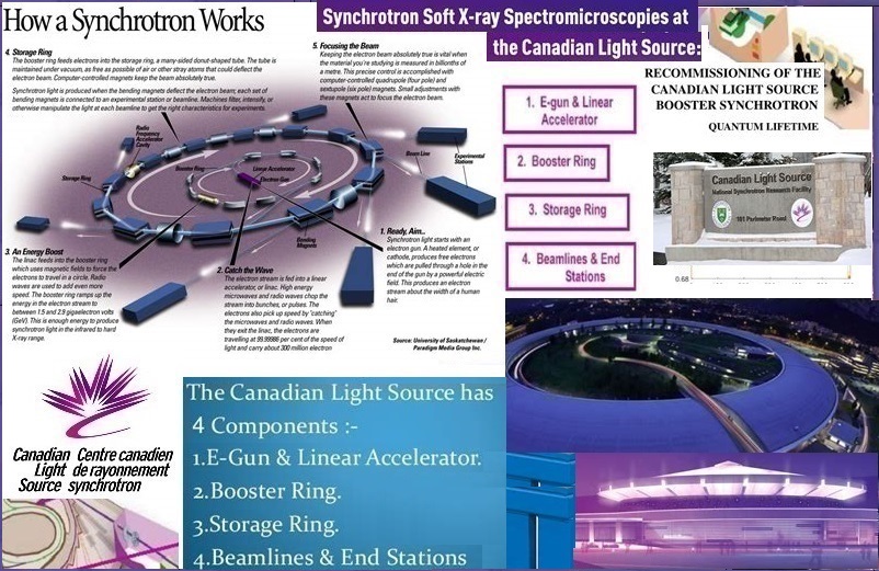
The Canadian Light Source Synchrotron (CLS) operates from the University of Saskatchewan as an international synchrotron facility. With more than one thousand users that represent scientists from all provinces in Canada and globally from twenty other countries research has focused from viruses to superconductors to dinosaurs. Since 2005, two thousand four hundred and sixteen Canadian Synchrotron Radiation Facility (CSRF) research papers has been published (eighty-five per cent were from Canadian researchers and the rest from external users). The CLS contains twenty-two beamlines that support a large selection of research presentations. Each beamline offers an exclusive spectral range that represents valuable knowledge. Scientists explore beamline specifications by their scientific identifications, spectral ranges, and specific techniques prior to research proposals.
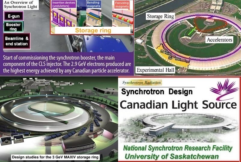
The history of the Canadian Light Source Synchrotron begins with the decision of its location. The Canada Foundation for Innovation (CFI) and the Atomic Energy of Canada Limited (AECL) collaborated until they had to decide between two university campuses – the Universities of Saskatchewan (US) and the University of Western Ontario (UWO). The University of Saskatchewan already had a veterinary vaccine laboratory and the infrastructure for expansion. CLS Saskatchewan Accelerator Laboratory (SAL) was funded by universities which included the University of British Columbia (UBC), Guelph University, and UWO. The Canadian Institute for Synchrotron Radiation (CISR) has expanded the facility’s beamlines as well as the building (each in two phases since its launch). Phase one of CLS was built with a 1.5 GeV (seven beamline) storage ring and full spectral ranges with superconducting bends to boost photon energies for soft X-rays. The beamlines included two infrared beamlines, three soft X-ray beamlines, and two hard X-ray beamlines. They served the purpose of synchrotron medical imaging for medical research groups. During phase two seven beamlines were added. The glass and steel expansion to accommodate the phase II medical imaging beamline (BMIT) also included the addition of other beamlines. In total five beamlines were added. To evaluate requests for beamtime, a committee was established under the chairmanship of Adam Hitchcock of McMaster University. In 2012, a high school group from La Loche Saskatchewan became the first to use the purpose-built educational beamline IDEAS. In the same year, CLS signed an agreement with the Advanced Photon Source synchrotron in the USA to allow Canadian researchers access to their facilities. The upgrade of the storage ring and to accommodate the Brockhouse beamline necessitated phase three. With further construction and expansion, CLS presently has a third-generation 2.9 GeV storage ring with longer straight sections which enables two insertion devices per straight and allows the ring to achieve currents of 100mA. An added feature of a storage beam is that a beamline can distribute two individual beamlines within one beam.
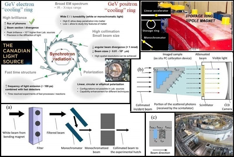
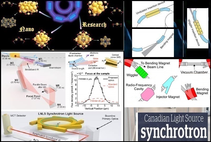
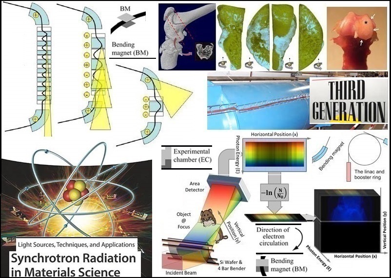
A synchrotron is one of the first accelerator concepts that enables the construction of large-scale facilities. Insertion devices are intermittent magnetic structures that are ‘inserted’ into accelerator tracks to arouse extremely brilliant, forward-directed synchrotron radiation emissions by forcing a stored charged particle beam to complete wiggles, or undulations, as they pass through the device. Electrons are accelerated by a synchrotron and injected into a storage ring in which they circulate before they produce synchrotron radiation. Beamlines originate at bending magnets or insertion devices. A high photon flux in a small area is a general requirement of a beamline. The electrons are captured by beamlines and end in experimental focal points that are used for experiments in chemistry, life science, materials science, molecular biology, and particle physics; also, for irradiation tests or to produce isotopes.
Spectrophotometers measure electromagnetic radiation (EMR) by dividing it into spectral ranges depending on each colour’s wavelength. Spectral wavelengths range from 10 nm hard gamma radiation wavelengths to kilometre long-wave broadcast wavelengths. Every colour exists in a wavelength range of 380-780 nm, which are the same wavelengths observed by the human eye. Spectrophotometry is a type of electromagnetic spectroscopy that controls quantitative measurements of reflective and transmissive properties; it is important in biology, chemical engineering, chemistry, material science, molecular biology, and physics. The food, forensic, and pharmaceutical industries regularly use spectrophotometry to test the colour and quality of their products with quantitative wavelength measurements. A spectrophotometer can measure spectral range and visible light spectrum wavelengths in forensic samples with insight and accuracy. A storage ring is a singular synchrotron (circular particle accelerator) in which a particle beam circulates to keep the kinetic energy of the particles constant. Storage of a particle depends upon the mass, momentum, and the charge of the particle. Storage rings store electrons, positrons, and protons; mostly electrons that radiate synchrotron radiation. In physics, EMR contains waves of the electromagnetic field that spread through space and transmit momentum and electromagnetic (EM) radiant energy which includes gamma rays, infrared, microwaves, radio waves, ultraviolet, visible light, and X-rays. These waves form part of the EM spectrum. EMR contains electromagnetic waves that are synchronised alternations of electric and magnetic fields. Depending on the frequency of alternations, different wavelengths of the spectrum are formed. In a vacuum, EM waves travel at the speed of light and are discharged by electrically charged particles that tolerate acceleration. For scientific and technical purposes, a synchrotron light source is electromagnetic radiation produced by a storage ring.
The Canadian Light Source houses 22 beamlines that support a wide selection of research applications. Each beamline offers a unique spectral range. The Quantum Materials Spectroscopy Centre (QMSC) beamline uses a unique dual-undulator method to investigate important materials in condensed matter physics. QMSC hosts two end stations designed for angle-resolved photoemission (ARPES) and spin-resolved photoemission (SARPES) experiments related to the motion of electrons in materials. QMSC was designed to control a broad array of energies to obtain vast information about electrons. This is a sixteen million Canadian dollar national effort funded by CFI for the construction of a state-of-the-art beamline facility committed to the performance of spin and angle-resolved photoemission spectroscopy (S+ARPES). The QMSC beamline’s spectral range is 15-1200 eV. The biomedical imaging and therapy (BMIT) beamlines are composed of a low-energy bending-magnet beamline (BMIT-BM) and a high-energy superconducting wiggler beamline (BMIT-ID). The bend magnet beamline is used to experiment with new imaging and therapy ideas and to validate techniques that can eventually be tested on the insertion device beamline. The two beamlines are dedicated to the imaging of biological tissues and radiation therapy research. New instruments are applied for general-purpose X-ray microtomography and imaging of fast processes. The bending-magnet and insertion device beamlines have been successful in their mission to image biological tissue and conduct live animal imaging studies. The BMIT laboratory will continue to provide synchrotron-specific imaging and therapy capabilities. Compared to global biomedical facilities, the BMIT facility implements high load systems for both BM and ID beamlines to address unsolved problems in medicine (human and animal), agriculture, and other biomedical sciences. The BMIT-BM beamline generates X-rays in the energy range of 12.6-40 keV for the imaging of lightweight, small samples and of little animals such as mice. The BMIT-ID wiggler-based beamline delivers a photon beam with adequate flux density to develop a monochromatic CT dataset in five to ten minutes (as fast as thirty seconds – depending on the subject matter). The spectral range spreads from 25 to 150 keV. The Canadian Nuclear Laboratories (CNL) is Canada’s premier nuclear science and technology organisation. When the National Research Universal nuclear reactor at Chalk River Laboratories in Ontario ceased production, an alternative supply of electron LINAC to produce the medical isotopes 99Mo (molybdenum-99) and 99mTc (technetium-99m) had to be found. CLS received fourteen million Canadian dollars in funding to install 35MeV LINAC in an abandoned underground experimental hall (a Saskatchewan Accelerator Laboratory formerly used for photonuclear experiments). Irradiation results are assessed by Winnipeg Health Sciences Centre. The Soft X-ray Microcharacterisation (SXRMB) beamline is a medium energy x-ray beamline which supports research from the biological, environmental, and chemical sciences. X-ray Photoelectron Spectroscopy (XPS) is a familiar laboratory technique to analyse the surface chemistry of samples. XPS is element-specific and can be used to track changes in chemistry (like oxidation state) of elements that are close to the surface (within a few nanometers). The SXRMB beamline has four end stations. Firstly, the XAFS end station and secondly, the Microprobe end station (the third Ambient Table and fourth High Energy XPS end stations are part of the Microprobe end station). XAFS end station with Bending Magnet – this end station can perform bulk analysis of solid samples like pellets and powders. Samples are placed under vacuum to optimise flux for lower energy edges such as phosphorus, silicon, and sulphur. This end station has fluorescence and total electron yield detection. Microprobe end station with Bending Magnet (K-B Mirro Sample Stage) – this end station provides a 10 × 10 μm beamspot to map experiments. Ambient Table – This multi-use end station is designed for bulk analysis of various samples. The end station can be configured for liquid and solid in-situ experiments. High Energy XPS – By setting the beamline to variable energies, XPS can be achieved at separate depths of the same sample, supplying information on superficial and mass properties of material. During May 2019, the XES spectrometer (easyXAFS) was installed into an atmospheric glovebox and the entire system was successfully integrated into the SXRMB beamline. The SXRMB beamline’s spectral range is 1.7 – 10 keV.
The Canadian Macromolecular Crystallography Facility (CMCF) consists of two beamlines, CMCF-BM and CMCF-ID, dedicated to crystallography. Both beamlines enable high-resolution structural studies of proteins, nucleic acids, and other macromolecules, satisfying the requirements of the most challenging and diverse crystallographic experiments. After the first experiments were conducted in 2006, the facility has seen a sharp increase in usage and has produced a substantial amount of data for the Canadian crystallographic community. The scientific aim of the CMCF-ID is to run a protein crystallography beamline suitable to study small crystals and crystals with large unit cells. In May 2022, a substantial upgrade to the beamline increased the overall flux, enabled micro-focus beam sizes of 5 µm, and greatly improved sample data. The two beamlines both have normal and high flux spectral ranges. The CMCF-BM beamline’s normal flux (DCM) range is 6 – 18 keV and its high flux (DMM) range is 7 – 10.5 keV (the 7 keV can reach a maximum flux of 8.157 keV). This mode is mostly used for routine screening and data collection. The CMCF-ID beamline’s normal flux (DCM) range is 5.0-20.0 keV and its high flux (DMM) range is 7.2-10.4 keV. Gathering of data using the high-flux DMM mode before the full dataset is collected presents a high risk of sample damage. The Resonant Elastic and Inelastic X-ray Scattering – Optical Layout Hi-Resolution Image (REIXS) beamline is a soft x-ray scattering facility for elastic and inelastic x-ray scattering experiments. The REIXS beamline, one of the top X-ray scattering beamlines in the world in quantum materials research, was funded by the Canada Foundation for Innovation. Using the X-ray Spectro microscopy beamline, a research team (led by scientists from the University of New York) created images of graphene that demonstrate how folds and ripples affect the conductivity of electrons. The REIXS beamline has a photon energy range from 95 eV – 2000 eV. The Brockhouse X-ray diffraction and scattering (BXDS) beamline for structural classification of many types of materials includes crystals, liquids, solids, and nanostructures under atmospheric conditions and at extreme pressures, temperatures, and magnetic fields. The Brockhouse Diffraction Sector is a suite of three beamlines (BXDS-WLE, BXDS-WHE, and BXDS-IVU) that provide a full range of diffraction and scattering techniques to describe the structure of materials. The spectral range of BXDS-WLE (Low Energy Wiggler Beamline) is 7-22 keV, of BXDS-WHE (High Energy Wiggler Beamline) is 20-94 keV, and of BXDS-IVU (Undulator Beamline) is 5-24 keV.
The Very sensitive Elemental and Structural Probe Employing Radiation from a Synchrotron (VESPERS) is a hard X-ray microprobe beamline able to provide a high level of analytical and structural information. X-ray diffraction, X-ray fluorescence spectroscopy, and X-ray absorption spectroscopy are to analyse a microscopic volume in the sample. Multi-bandpass and pink beam ability are built-in, and a mm-sized beam is also available. The VESPERS beamline’s spectral range is 6 – 30 keV. The BioXAS-Spectroscopy beamline is a group of two independently operating end stations that share a wiggler source. Spectroscopy is designed to measure the XAS spectra of transition elements in bulk biological samples with diluted concentrations. It is a multi-resolution high-sensitivity X-ray fluorescence (XRF) imaging beamline with capabilities to collect XRF imaging data in macro-, micro-, and nano modes. The sector was designed to support health and life sciences as well as environmental research. Among other organisations, the beamline was funded by the Canada Foundation for Innovation and the Government of Saskatchewan. The BioXAS spectral range is 5-32 keV. The Hard X-ray Micro-Analysis (HXMA) beamline is used to study the fundamental local structure environment of materials in-situ and ex-situ, accepting bulk and thin film samples). The HXMA beamline’s spectral range is 5 – 40 keV. The Spherical Grating Monochromator (SGM) beamline is designed for soft x-ray absorption spectroscopy and x-ray photoelectron spectroscopy with the importance on environmental samples and catalytic materials. A readily accessible end station with four silicon drift detectors is an ideal model for in-situ and in-operando experiments. The beamline is fitted with two in-line end stations for high resolution x-ray absorption spectroscopy and photoelectron spectroscopy. The SGM beamline’s spectral range is 250-2000 eV. The X-ray Synchrotron Radiation (XSR) and Optical Synchrotron Radiation (OSR) beamlines are two independent diagnostic beamlines. The Soft X-ray Spectromicroscopy (SM) beamline supports research programs such as biological applications, environmental science, magnetic imaging, and polymer science. The SM beamline has a photon energy range from 130 eV to 3000 eV. The Synchrotron Laboratory for Micro and Nano Devices (SyLMAND) is an x-ray beamline and pre- and post-processing laboratory in a cleanroom environment. The SyLMAND beamline’s spectral range is 1 – 15 keV. The Variable Line Spacing Plane Grating Monochromator (VLS-PGM) beamline is fitted with four VLS gratings to cover the designed energy range of the high resolution, low energy spectroscopic beamline which enables research in materials of fundamental and applied nature. The VLS-PGM beamline’s spectral range is 15-250 eV.
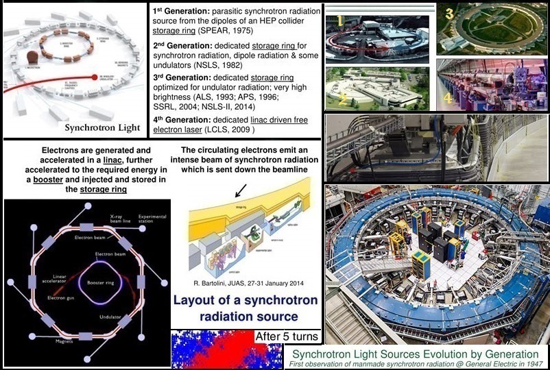
The Industry Development Education Applications Students (IDEAS) beamline is the instant access beamline for industry, in-house science, education programs, and to support the development and testing of beamline equipment and software. IDEAS is not available to other programs or researchers. The beamline’s spectral range is 2.2 – 13.4 keV. CLS is known for its industrial science and high school education programs. Their education program ‘Students on the Beamlines’ is funded by NSERC Promoscience. The program allows high school students to experience using the CLS beamlines and to observe the work of a scientist. It also gives them the opportunity to develop active research (a very rare phenomena in schools) and offers them direct access to the use of a particle accelerator (which is even rarer). Students from six provinces as well as the Northwest Territories have been directly involved in experiments on the IDEAS beamline; some have produced publishable research. In 2012, CLS was awarded the Canadian Nuclear Society’s Education and Communication Award for its commitment to community outreach, increased public awareness of synchrotron science, and innovative secondary educational programs such as Students on the Beamlines. The Mid Infrared Spectromicroscopy (Mid IR) offers a state-of-the-art Fourier Transform IR spectrometer and microscope to deliver diffraction-limited spatial resolution to an increasing sequence of experimental projects. The benefits of high brightness infrared synchrotron light are motivating researchers to revisit existing techniques and explore new experiments. The spectral range of Mid IR is 560 – 6000 cm-1 The Far Infrared Spectroscopy (Far IR) provides infrared light 100 – 1000 times brighter than standard laboratory sources, enabling the spectroscopic study of molecules with considerably higher precision and sensitivity. The spectral range of Far IR is 5 – 1200 cm-1 (synchrotron source) and 30 – 5000 cm-1 (conventional sources). The storage ring has twelve straight sections (ss) with induction devices, wigglers, and undulators to increase the beam energy. Ss1 injection straight, ss2 electron beam diagnostic straight, ss3 reserved for future use, ss4 chicaned: in-vacuum undulator, in-vacuum wiggler, ss5 superconducting wiggler, ss6 superconducting wiggler, ss7 chicaned: in-vacuum undulator, out-vacuum wiggler, ss8 in-vacuum undulator, ss9 single length dual period undulator, ss10 chicaned: undulators, ss11 chicaned: undulators, and ss12 RF cavity. The upstream accelerator complex is a 220 keV DC thermionic RF gun. A six section LINAC increases the beam energy from 220 keV to 250 MeV. An energy compression system (3 magnet chicane and RF cavity) compresses the beam energy spread by a factor of ten. The beam is transported into a booster ring and ramped up from 250 MeV to 2.9 GeV.
An international team (led by the University of Calgary professor Ken Ng) solved the structure of RNA polymerase using X-ray crystallography. This enzyme replicates itself as the Norwalk virus as it spreads through the body. It has been linked to other super viruses such as the common cold, hepatitis C, and West Nile virus; its replication is liable for the onset of such viruses. CLS scientist (Luca Quaroni) and the University of Saskatchewan professor (Alan Casson) identified biomarkers inside individual cells with infrared microscopy from tissue related to Barrett’s oesophagus. This disease can lead to the aggressive cancer known as oesophageal adenocarcinoma. Research was done by the University of Saskatchewan on peptides and by UWO on materials for organic light-emitting diodes. Researchers from Lakehead University and the University of Saskatchewan investigated the deaths of Royal Navy sailors buried in Antigua in the late 1700s. With the use of X-ray fluorescence, they looked for trace elements such as lead and strontium in bones from the excavated naval cemetery. The University of Regina and the Royal Saskatchewan Museum examined dinosaur fossils after discovering a Tyrannosaurus in Saskatchewan during 1991, one of the largest and most complete T-rex skeletons ever found. To study the impact of the environment on such animals, they explored the concentration of elements in the skeleton’s bones.
To access the synchrotron facility interested researchers are required to apply through a peer review system that verifies the quality of the proposed science experiments, regardless of academic, governmental, industrial, national, or regional relations. CLS has an industrial group within the facility who are represented by industrial liaison scientists who make synchrotron procedures available to non-synchrotron experts. An economic impact study revealed that the Canadian Light Source Incorporated added forty-five million Canadian dollars to Canada’s annual gross domestic product for the two financial years 2010 and 2011.